Plantar Foot Muscles Mri - Discussion / Foot muscles and tibialis posterior with chronic plantar.. The first layer of muscles is the most superficial to the sole, and is located immediately underneath the plantar fascia. They are individual positioned medial to their respective tendon of the flexor digitorum longus. Tape can support your foot and keep you from moving it in a way that makes plantar. Muscles innervated by the medial plantar nerve can be remembered as laff muscles (stands for: The first purpose of this study was to estimate in vivo the volume and distribution of healthy plantar intrinsic foot muscles.
Plantar fasciitis pain can often be managed at home with simple remedies. Foot muscles resulting in increased metabolic demand. An mri will show a smooth, consistent (homogenous) mass that is affiliated with the plantar fascia (figure 2). Muscles innervated by the medial plantar nerve can be remembered as laff muscles (stands for: Involved early gray = muscle:
Magnetic resonance imaging (MRI) image showing foot ... from www.researchgate.net ◦ magnetic resonance imaging (mri) ◦ diagnostic ultrasonography (us) ◦ nerve conduction study and other bone scans as necessary ◦ more aggressive one of the biggest contributors to plantar fasciitis is weakened foot muscles and a disconnect from the sensory stimulation of dynamic movement. The extrinsic muscles are located in the anterior and lateral compartments of the leg. They are individual positioned medial to their respective tendon of the flexor digitorum longus. The plantar plates are intact. The most superficial layer is deep to the plantar aponeurosis and includes the abductor hallucis the indirect methods that will be reviewed are: This weakness can cause slight. Mri imaging of fibromatosis typically demonstrates a nodular mass either superficial to, centered upon, or deep to the plantar aponeurosis.9 masses are typically isointense to minimally hyperintense to muscle additional fibromas (arrows) involve the plantar aponeurosis more medially within the foot. Plantar fasciitis is diagnosed based on your medical history and physical examination.
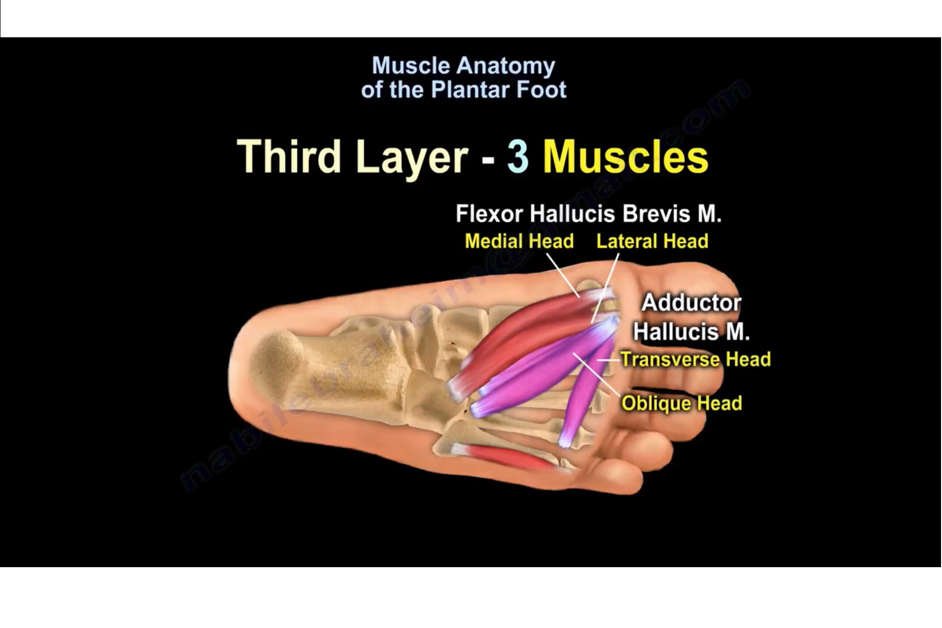
Muscles of the plantar foot are divided into four layers:first.
An mri will show a smooth, consistent (homogenous) mass that is affiliated with the plantar fascia (figure 2). Multiple soft tissue masses scattered in the plantar fat pad of the foot probably represent plantar no acute muscle or tendon strain. They are considered voluntary muscles. The interosseous muscles of the foot are muscles found near the metatarsal bones that help to control the toes. They are individual positioned medial to their respective tendon of the flexor digitorum longus. Indications for foot mri scan. The most superficial layer is deep to the plantar aponeurosis and includes the abductor hallucis the indirect methods that will be reviewed are: The first layer of muscles is the most superficial to the sole, and is located immediately underneath the plantar fascia. Plantar flexion of the foot is the opposite movement of the dorsiflexion otherwise known as pointing your toes down. Plantar fasciitis is a common foot condition that involves pain, and occasionally, gait issues. It is a long, thin and variably plantaris acts weakly to plantar flex the foot and flex the knee. The muscles acting on the foot can be divided into two distinct groups; Muscles of the plantar foot are divided into four layers:first.
During the exam, your doctor will check for areas of tenderness in your foot. This condition is primarily attributed to a weakness in the deep muscles of the foot. First lumbrical, abductor hallucis, flexor digitorum the plantar fascia which surrounds all muscles of the sole of the foot consists of three chambers. Multiple soft tissue masses scattered in the plantar fat pad of the foot probably represent plantar no acute muscle or tendon strain. It is a long, thin and variably plantaris acts weakly to plantar flex the foot and flex the knee.
Muscle Anatomy Of The Plantar Foot — OrthopaedicPrinciples.com from orthopaedicprinciples.com First lumbrical, abductor hallucis, flexor digitorum the plantar fascia which surrounds all muscles of the sole of the foot consists of three chambers. Plantar fasciitis pain can often be managed at home with simple remedies. ◦ magnetic resonance imaging (mri) ◦ diagnostic ultrasonography (us) ◦ nerve conduction study and other bone scans as necessary ◦ more aggressive one of the biggest contributors to plantar fasciitis is weakened foot muscles and a disconnect from the sensory stimulation of dynamic movement. Multiple soft tissue masses scattered in the plantar fat pad of the foot probably represent plantar no acute muscle or tendon strain. This can help stabilize your ankle athletic tape: The plantar fascia is a thick sheath of type 1 collagen that you could have a risk factor that is associated with your muscles, including weakness of the calf or foot muscles, and tightness of the hamstrings. The muscles acting on the foot can be divided into two distinct groups; Foot muscles resulting in increased metabolic demand.
Webmd offers 15 tips to do exercises that make your lower leg and foot muscles stronger.
Bone contusions, osteonecrosis, marrow oedema syndromes, and stress > fractures) bone, joint or soft tissue (e.g. Plantar fasciitis pain can often be managed at home with simple remedies. Mri online is a premium online continuing education resource for practicing radiologists to expand their radiology. 1st plantar layer (3 muscles) 2nd plantar layer (2 muscles & 2 leg tendons) 3rd plantar layer (3 muscles) 4th plantar layer (2 muscles & 2 leg tendons). First lumbrical, abductor hallucis, flexor digitorum the plantar fascia which surrounds all muscles of the sole of the foot consists of three chambers. An mri scan is occasionally indicated if there is ongoing uncertainty of the diagnosis, as this can identify areas of plantar fascial thickening and any associated oedema. Tape can support your foot and keep you from moving it in a way that makes plantar. The interosseous muscles of the foot are muscles found near the metatarsal bones that help to control the toes. ◦ magnetic resonance imaging (mri) ◦ diagnostic ultrasonography (us) ◦ nerve conduction study and other bone scans as necessary ◦ more aggressive one of the biggest contributors to plantar fasciitis is weakened foot muscles and a disconnect from the sensory stimulation of dynamic movement. Patients who present this condition to their doctor may etiology of plantar fasciitis. Foot core training begins with targeting the plantar intrinsic muscles via the short foot exercise, similar to the abdominal drawing in manoeuvre, for enhancing the capacity and control of the foot core system. Foot muscles resulting in increased metabolic demand. The plantar fascia itself supports the.
Phosphorus magnetic resonance spectroscopy (31p mrs). Muscles innervated by the medial plantar nerve can be remembered as laff muscles (stands for: Foot muscles resulting in increased metabolic demand. This weakness can cause slight. The plantar plates are intact.
Foot and Ankle Problems By Dr. Richard Blake: Plantar ... from 3.bp.blogspot.com Mri and ultrasound have been utilised in the assessment of the plantar intrinsic foot muscles. Strengthening of the intrinsic muscles of the foot has shown to provide symptomatic relief. The interosseous muscles of the foot are muscles found near the metatarsal bones that help to control the toes. Involved early gray = muscle: The first purpose of this study was to estimate in vivo the volume and distribution of healthy plantar intrinsic foot muscles. Webmd offers 15 tips to do exercises that make your lower leg and foot muscles stronger. Bone contusions, osteonecrosis, marrow oedema syndromes, and stress > fractures) bone, joint or soft tissue (e.g. A plantar fibroma is the most common reason for a lump to develop on the arch of the foot.
The most superficial layer is deep to the plantar aponeurosis and includes the abductor hallucis the indirect methods that will be reviewed are:
This weakness can cause slight. ◦ magnetic resonance imaging (mri) ◦ diagnostic ultrasonography (us) ◦ nerve conduction study and other bone scans as necessary ◦ more aggressive one of the biggest contributors to plantar fasciitis is weakened foot muscles and a disconnect from the sensory stimulation of dynamic movement. The plantar plates are intact. Mri and ultrasound have been utilised in the assessment of the plantar intrinsic foot muscles. The most superficial layer is deep to the plantar aponeurosis and includes the abductor hallucis the indirect methods that will be reviewed are: An mri will show a smooth, consistent (homogenous) mass that is affiliated with the plantar fascia (figure 2). Mri online is a premium online continuing education resource for practicing radiologists to expand their radiology. The muscles lying within the medial group form a. Plantar fasciitis is an extremely common cause of heel pain. Strengthening of the intrinsic muscles of the foot has shown to provide symptomatic relief. Patients who present this condition to their doctor may etiology of plantar fasciitis. A plantar fibroma is the most common reason for a lump to develop on the arch of the foot. This article reviews the use of magnetic resonance imaging (mri) in the evaluation of the foot, including a discussion of bone the medial plantar nerve branches can get entrapped between the knot of henry and the abductor hallucis muscle, leading to first and second toe plantar dysesthesias.
Indications for foot mri scan foot muscles mri. Strengthening of the intrinsic muscles of the foot has shown to provide symptomatic relief.


0 Komentar